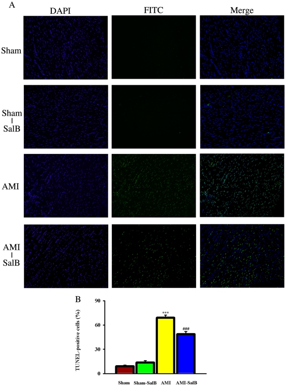
Figure 6. SalB protected cardiomyocytes from apoptosis detected by TUNEL assay in ischemic area of heart.
(A) Representative pictures of each treatments. The total cells were identified by DAPI stain, and the positive apoptosis cells were identified by FITC stain. (B) Quantitative data of apoptosis cells. ***p<0.001 compared with Sham, ###p<0.001 compared with AMI. n = 10 for each group. At least three time experiments were repeated and the representative figures were shown.

