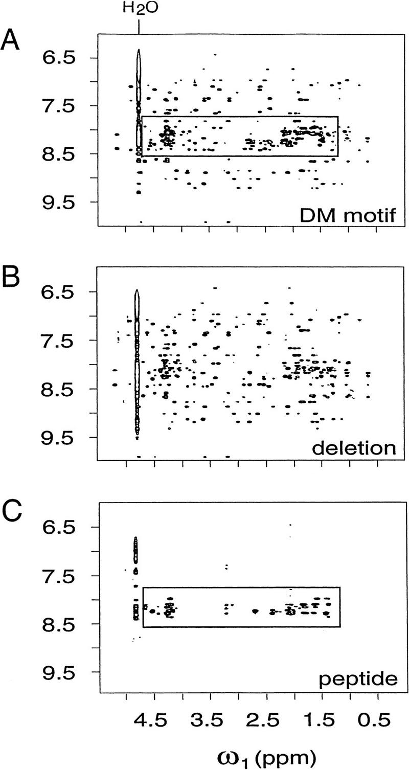
Figure 5.
NOEsY spectra demonstrate “peptide dissection” of intact DM motif (A) into major and minor fragments: Zn-binding module (DMΔ; B) and carboxy-terminal peptide (peptide DM-p; C). Shown are respective spectra in H2O at 25°C and 600 MHz; mixing times were in each case 175 msec. Cross peaks from amide and aromatic protons (vertical scale, θ2) and aliphatic protons (horizontal scale, θ1) are shown. Boxed regions highlight unresolved envelope in intact DM domain (A) assigned to nascent carboxy-terminal helix (C); this feature is absent in spectrum of DMΔ (B). Peptides were made 1.5 mm in 50 mm deuterated Tris-HCl (pH 6.5) in 90% H2O and 10% D2O. Spectra A and B also were obtained in presence of 4 mm ZnCl2.

