
Figure 6.
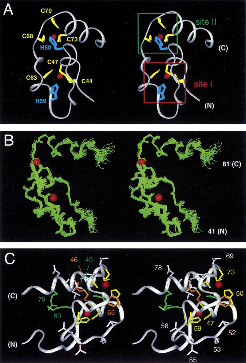
(A) Stereo ribbon representation of the solution structure of the DM domain (DSX residues 41–81). An ensemble of side chains of the coordinating cysteate (yellow) and histidine (blue) side chains and two bound Zn2+ atoms (red) are shown. The two intertwined zinc-binding sites are designated sites I and II (boxes at right). (B) Ensemble of main chain structures (stereo pair) aligned according to the main chain atoms of residues 41–81. The positions of Zn2+ atoms are shown by red spheres (50% of van der Waal radius). (C) Structural relationships among side chains of conserved (green), otherwise ordered (white), or invariant ligand-binding (green) residues. Ligand-binding residues are C44, C47, H59, C63 and H50, C68, C70, C73 (see Fig. 1B). Conserved residues N49 and R79 are shown in green whereas side chains of R46 and F65 are shown in gold to highlight structural environments (see text). Otherwise well-order residues include side chains of N43, A45, R48, L52, K53, T55, L56, Y67, T69, L75, T76, A77, and D78; criterion for inclusion is an ensemble side chain RMSD of <1.0 Å.