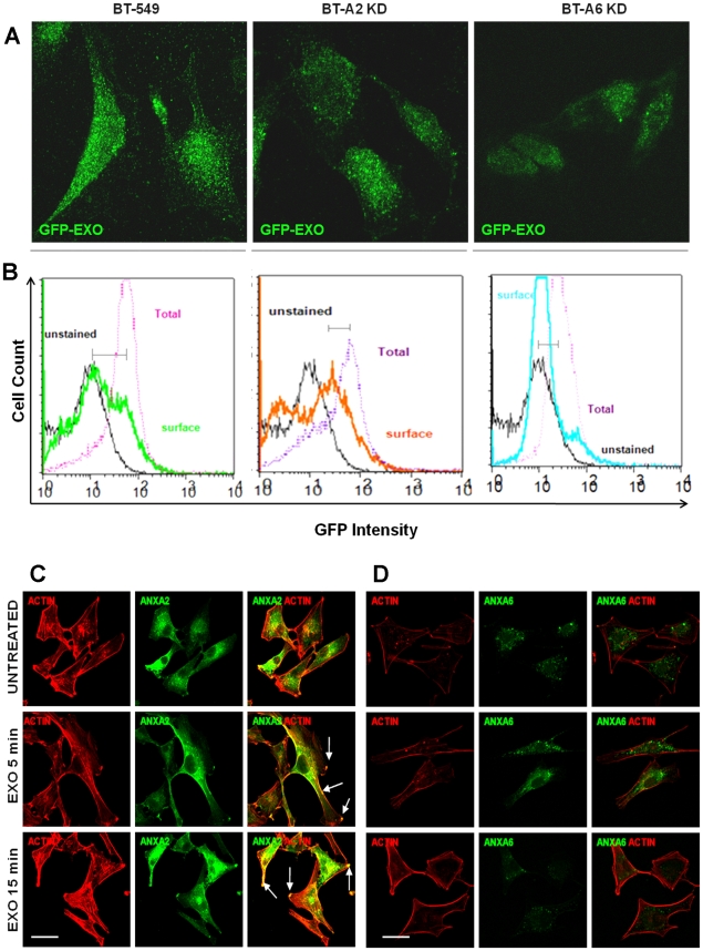
Figure 8. Annexin-mediated uptake and disposition of exosomes on the cell surface.
Parental BT-549, BT-A2-sh and BT-A6-sh were grown on glass coverslips or 10 cm dishes and incubated with GFP-CD63 exosomes and analyzed by (A) confocal microscopy or (B) Flow Cytometry. In panels C and D, BT549 cells were grown on glass cover-slips, serum starved for 24 hours washed and stimulated without (control) and with 100 µg/mL exosomes in HBSS 0.5% BSA 1 mM Ca2+ 1 mM Mg2+ for 5 and 15 minutes at 37°C and fixed with PFA. Cells were then treated with rhodamine phalloidin for actin staining followed by antibodies to AnxA2 (C) and AnxA6 (D). Images were acquired with a Nikon TE2000 confocal microscope. Bar is 20 µm.

