Figure 3.
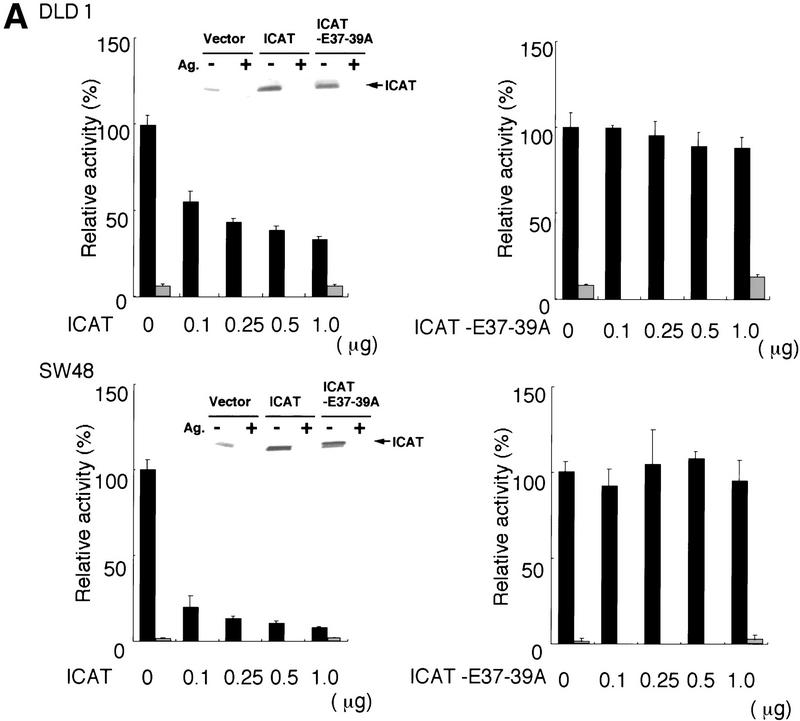
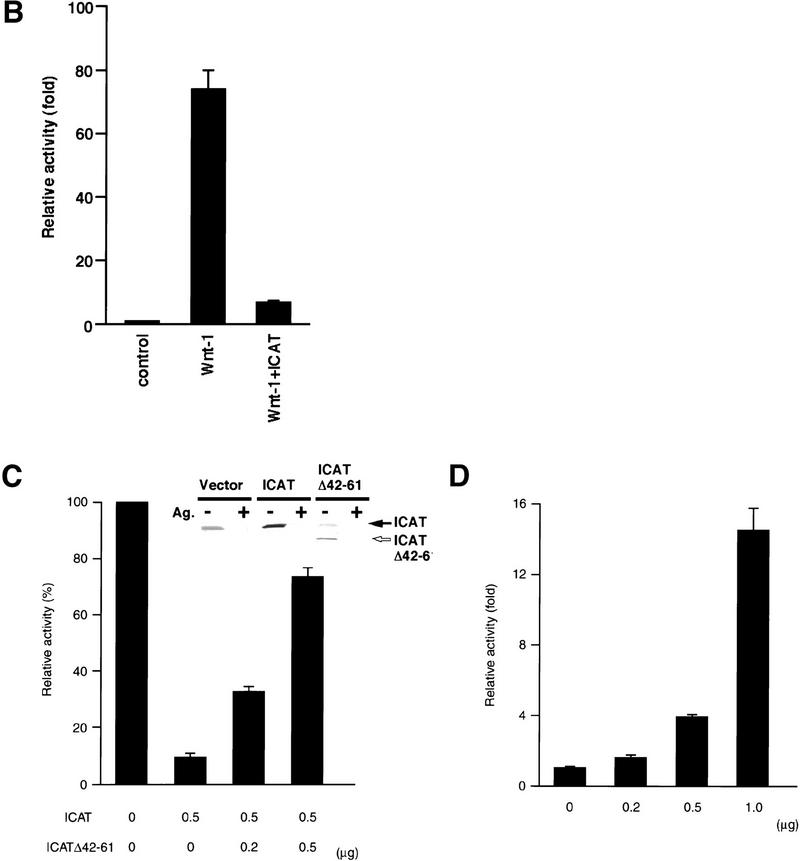
Effect of ICAT on β-catenin–TCF-regulated transcription. (A) ICAT represses constitutively activated β-catenin–TCF-mediated transcription in colorectal cancer cells. The human colorectal cancer cell lines DLD-1 and SW48 were transfected with the luciferase reporter plasmid TOPtkLuciferase (solid bars) or FOPtkLuciferase (shaded bars), and increasing amounts of ICAT, ICAT–E37–39A, and luciferase activity were measured (15). TOPtkLuciferase contains optimal and FOPtkLuciferase contains mutated TCF-binding sites placed upstream of a luciferase reporter gene. Luciferase activities are expressed relative to samples containing no ICAT and ICAT–E37–39A. (Insets) Detection of endogenous ICAT and exogenously expressed ICAT and ICAT–E37–39A (indicated by closed arrow). Lysates prepared from DLD-1 or SQ48 cells transfected with the indicated plasmids (1.0 μg) were subjected to immunoprecipitation and subsequent immunoblotting with anti-ICAT antibodies. (Ag+) Antibodies were preincubated with antigen before use in immunnoprecipitation. (B) ICAT represses Wnt-1-induced β-catenin–TCF-mediated transactivation. 293 cells were transfected with luciferase reporter plasmids, ICAT, and Wnt-1, and luciferase activity was measured. (C) ICAT–Δ42–61 abrogates the inhibitory effect of wild-type ICAT on β-catenin–TCF-mediated transcription. 293 cells were transfected with the luciferase reporter (0.05 μg), β-catenin (0.01 μg), ICAT, and ICAT–Δ42–61, as indicated. (Inset) Detection of endogenous ICAT, exogenously expressed ICAT (indicated by solid arrow), and ICAT–Δ42–61 (open arrow). Lysates prepared from 293 cells transfected with the indicated plasmids (0.5 μg) were subjected to immunoprecipitation and subsequent immunoblotting with anti-ICAT antibodies. (Ag+) Antibodies were preincubated with antigen before use in immunoprecipitation. (D) ICAT–Δ42–61 activates endogeneous β-catenin–TCF-mediated transcription. 293 cells were transfected with the luciferase reproter (0.05 μg) and increasing amounts of ICAT–Δ42–61 (as indicated).


