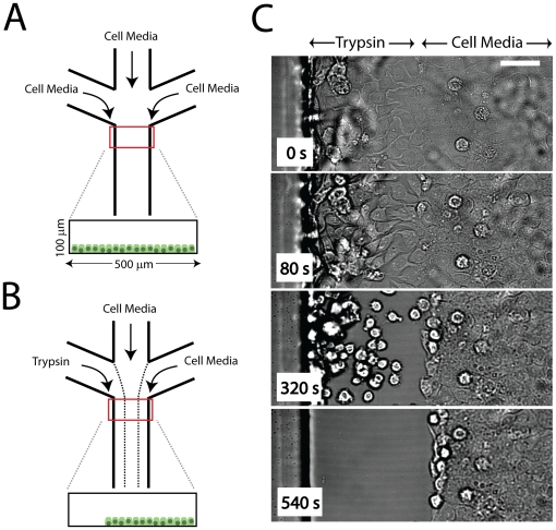
Figure 1. Experimental Design.
(A) Schematic of Experimental Setup. Three inlets supply cell media to an epithelial sheet cultured in the  wide,
wide,  high channels. This is done under no flow. (B) After cells have reached confluency, 15
  l/min flow (
 ) is applied, leading to three separated laminar streams. One stream contains 0.05% trypsin, while the other two contain cell media. This cleaves cells from the channel as shown in (C). On average, cleavage takes 5 min. Images are acquired in brightfield.

