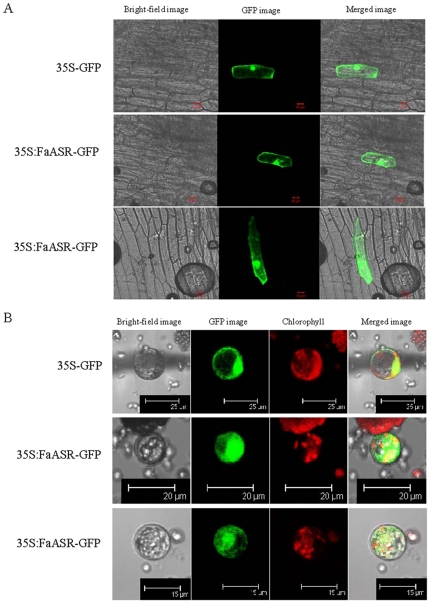
Figure 2. Subcellular localization of FaASR in onion epidermal cells (A) or tobacco protoplasts (B).
Constructs carrying GFP or FaASR-GFP were bombarded into onion epidermal cells or transfected into tobacco protoplasts as described in the Materials and methods. GFP and FaASR-GFP fusion proteins were transiently expressed under control of the CaMV 35S promoter and observed with a laser scanning confocal microscope. Experiments were done in triplicate resulting in the same fluorescence pattern, and two different images for FaASR-GFP were presented. The FaASR-GFP fusion protein was present in the cell outlines and the nuclear. The length of Bar was indicated in the photos.

