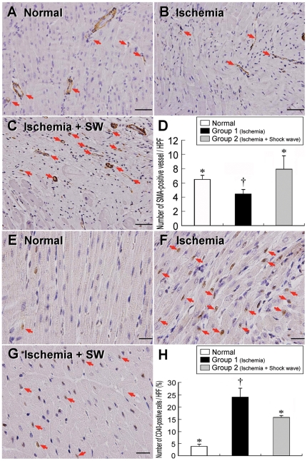
Figure 6. Immunohistochemical staining of α-SMA and CD40 nuclei in LV myocardium.
Upper panel: α-SMA immunohistochemical staining (200x) showing notably lower number of small vessel (red arrows) after ischemia (B) than in normal controls (A) and ischemia with shock wave treatment (C). †p<0.002 between the indicated groups (D). Scale bars in right lower corner represent 50 µm. Lower Panel: IHC staining (400x) showing the CD40-positively stained cells were remarkably higher after ischemia (F) than in normal controls (E) and ischemia with shock wave treatment (G). †p<0.001 between the indicated group (H). Scale bars in right lower corner represent 20 µm.

