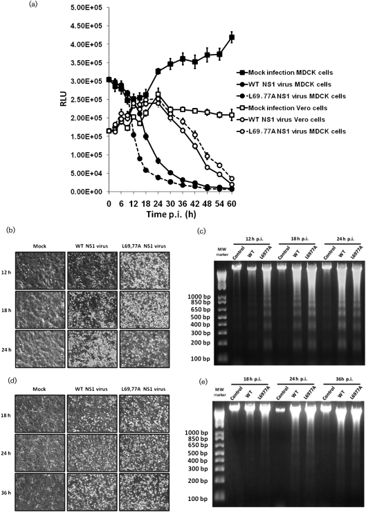
Fig. 3.
L69,77A NS1 virus infection induces more apoptosis in MDCK cells, but not in Vero cells, compared with the WT NS1 virus. (a) MDCK and Vero cells grown on 96-well plates were mock infected or infected with WT NS1 or L69,77A NS1 viruses at an m.o.i. of 5. Cell viability after infection, as reflected by relative luminescence unit (RLU), was determined by CellTiter-Glo assay at every 3 h until 18 h and every 6 h afterwards until 60 h. (b–e) MDCK and Vero cells grown on six-well plates were mock infected or infected with viruses at an m.o.i. of 5. At various time points (12, 18 and 24 h for MDCK cells and 18, 24 and 32 h for Vero cells) after infection, images were collected by a phase-contrast microscope (b and d in MDCK and Vero cells, respectively) and DNA fragmentation was analysed by 1.5 % agarose gels (c and e in MDCK and Vero cells, respectively).

