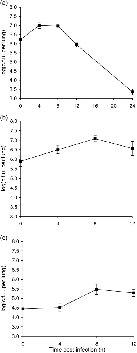
Fig. 1.
Growth of S. maltophilia strain K279a in the lungs of A/J mice. (a–c) Mice were intranasally inoculated with different amounts of strain K279a (▪), resulting in different levels of deposition (see t = 0, in a–c), and then at 4, 8, 12 and 24 h post-inoculation, the c.f.u. in total lung homogenates were determined by plating. Data are the mean±sd obtained from four or five infected animals. Significant increases in c.f.u. relative to the value at t = 0 were obtained at 4 and 8 h in (a) and (b) and at 8 and 12 h in (c) (Student's t test, P<0.05).

