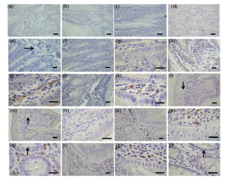
Fig. 1.
Immunohistochemical localizations of FoxO proteins in the duodenum of Sprague-Dawley rats
The immunohistochemical signals appear brown and the counterstaining background appears blue in color. Immunohistochemical localization of FoxO1: 21-d-old (b), 2-month-old (c), 6-month-old (d); Immunohistochemical localization of FoxO3a: 21-d-old (f, g), 2-month-old (h, i), 6-month-old (j, k); Immunohistochemical localization of FoxO4: 21-d-old (m, n), 2-month-old (o, p, q), 6-month-old (r, s, t). (q) and (t) are transverse sections. In control sections of FoxO1 (a), FoxO3a (e), and FoxO4 (l), normal albumin bovine V was used instead of primary antibody. →: muscularis mucosa; ↓: submucosa; ↑: intestinal gland. Bar=50 µm

