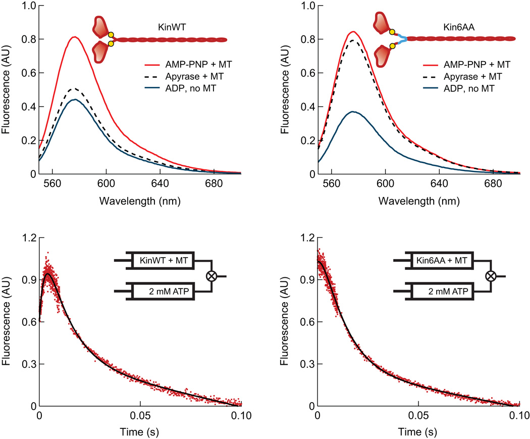
Figure 4.
Fluorescence data for Kin6AA and KinWT with TMR probes attached to both neck-linkers. (a, b) Steady-state TMR fluorescence emission spectra for KinWT (a) and Kin6AA (b), which monitors neck-linker separation under the following conditions: microtubules plus 2 mM AMP–PNP (red), microtubules plus apyrase to remove nucleotides (black, dashed), and 2 mM ADP without microtubules (blue). The large signal increase in the absence of nucleotide (apyrase present) for Kin6AA is consistent with neck-linker separation. In the inset cartoons, approximate locations of the TMR probes (at position 333) are indicated (yellow circles), as well as the neck-linker inserts (blue lines). (c, d) Pre-steady-state TMR kinetic records for KinWT and Kin6AA. TMR-labeled motors complexed to a five-fold excess of microtubules and treated with apyrase were mixed with 2 mM ATP. The initial increase in fluorescence seen for KinWT is absent for Kin6AA, indicating that prior to mixing with ATP, both heads of Kin6AA are bound to the microtubule, and consequently, the neck-linkers of this mutant are separated.

