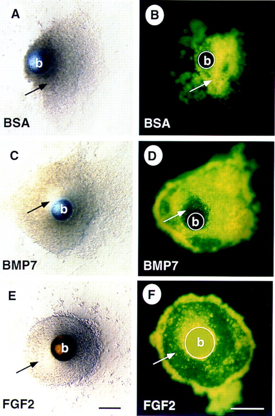
Figure 2.

BMP7 promotes survival of the nephrogenic mesenchyme. Mesenchyme was isolated from 11.5-dpc embryos and grown in culture for 48 hr in the presence of a bead containing BSA (A,B), BMP7 (C,D), or FGF2 (E,F). Explants treated with BSA appear granular and refractory to light. In contrast, there is a well organized, translucent region of mesenchyme in the vicinity of the BMP7-soaked bead (cf. cells indicated by arrow in A and C). Staining with the vital dye TOPRO-1 reveals that cells in the BSA-treated mesenchyme are dying as indicated by fluorescent signal (B), whereas, in the BMP7-treated mesenchyme, cells in the translucent region do not fluoresce, indicating that these cells are viable (cf. cells indicated by arrow in B and D). Interestingly, most of the cells in the explant survive when treated with FGF2 (E,F). Note that the heparin–Sepharose beads in E and F display autofluorescence. (b) Bead. Scale bar, 100μm.
