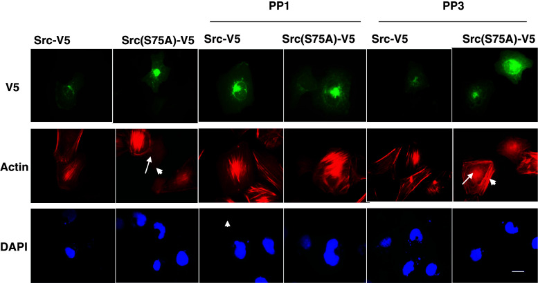
Fig. 4.
Src(S75A) reduces the formation of central stress fibers. Transfected cells expressing Src-V5 or Src(S75A)-V5 were replated on fibronectin-coated chamber slides for 1 h. Cells were then incubated for 2 h in absence or presence of PP1 or PP3. Representative fluorescence images of V5-expressing cells (upper images; green channel), phalloidin-stained actin cytoskeleton (middle images; red channel), and DAPI-stained nuclei (lower images; blue channel) are shown. Well-formed central stress fibers are present in 96.3% of Src-V5 cells at the selected fluorescence intensity. In contrast, 87.5% of cells transfected with Src(S75A)-V5 at comparable fluorescence intensity either showed no central polymerized actin (long arrow) or had peripheral actin fibers (short arrow). PP1 did not disturb central stress fiber formation in Src-V5 cells, but rescued the disrupting effect of Src(S75A)-V5 on central stress fibers. PP3 did not affect stress fiber organization either in Src-V5 or Src(S75A)-V5 cells (arrows). This experiment is representative of three experiments with similar results. Scale bar = 20 μm

