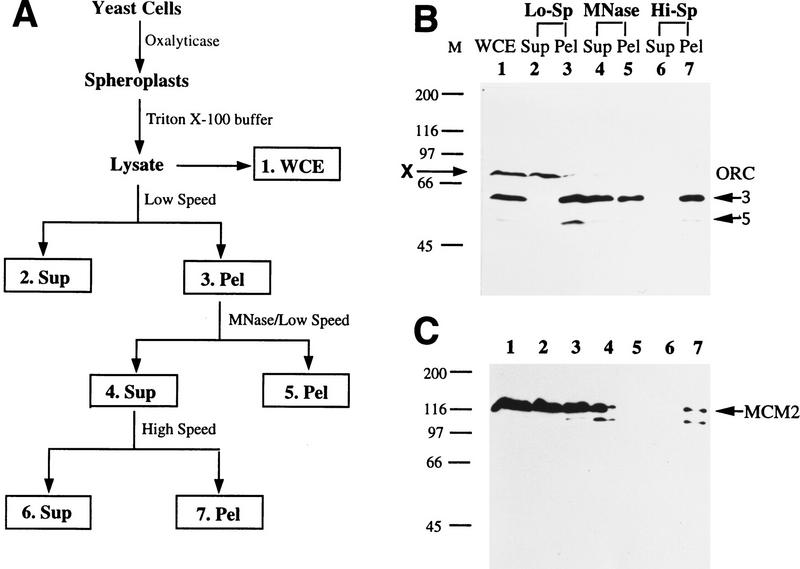
Figure 1.
A protein chromatin-binding assay. (A) Diagram of the chromatin-binding assay. Fractions are as follows. (1. WCE) whole cell extract from the total lysate; (2. Sup) first low speed supernatant containing nonchromatin-binding proteins; (3. Pel) crude chromatin pellet; (4. Sup) polynucleosome-containing supernatant after limited MNase digestion of the crude chromatin pellet; (5. Pel) remaining solid after MNase digestion; (6. Sup) supernatant from ultracentrifugation; (7. Pel) high-speed pellet containing polynucleosomes. (B) All seven fractions from the chromatin-binding assay (except that the first low-speed pellet was not washed) using asynchronous W303-1A cells were Western blotted for Orc3 and Orc5 subunits. The same cell equivalent was loaded in all seven fractions. (X) cross-reacting band by the Orc3p antibody. (C) The same blot used in B was probed for Mcm2p. The arrow points to the upper band of endogenous Mcm2p, which comigrates with purified, baculovirus-expressed Mcm2p from sf9 cells (data not shown, but see Fig. 2F). The lower band is likely degradation product of Mcm2p.

