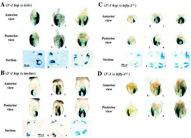
Figure 7.
Response of the 3′-1hsp construct to iv, inv, and lefty-1 mutations. Expression of the 3′-1hsp transgene was examined in iv/iv (A), inv/inv (B), and lefty-1−/− (C) mutant embryos. Anterior and posterior views, as well as a transverse section, are shown for each transgenic embryo. In iv/iv embryos (A), the staining in the LPM and PFP was either left-sided, right-sided or bilateral. In inv/inv embryos (B), the staining in the LPM and PFP was right-sided (embryo at left) or bilateral (embryos at middle and right). In lefty-1−/− embryos, the staining in the LPM and PFP was initially normal (embryo at left). At later stages (embryos at middle and right), however, the staining appeared on the right LPM. The staining in the PFP, increased and became bilateral (C). In lefty-1−/− embryos harboring 3′-1 transgene (D), X-gal staining in the PFP, which was not detected in the wild-type embryos (Fig. 4A), became apparent. X-gal staining pattern in LPM was similar to that in C. lacZ expression in the PFP always preceded that in the right LPM.

