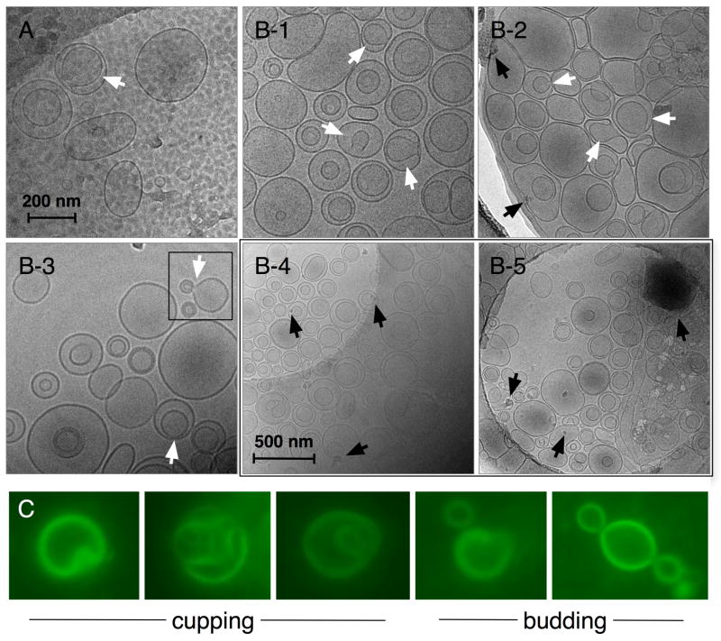
Figure 2.
Cryo-TEM micrographs of (A) FTLs without encapsulated DOX and (B-1 to B-5) FTMLs containing encapsulated DOX and SPIO nanoparticles. Both samples were prepared by thin film hydration in 10× PBS (pH 7.4) followed by membrane extrusion at 200 nm. The 200 nm scale bar is common to A and B-1 to B-3, and the 500 nm scale bare is common to B-4 and B-5. White arrows denote cup-shaped liposomes containing a visible pore or mouth and black arrows denote SPIO nanoparticles and nanoparticle aggregates. The square region denotes liposome budding. Representative magnified fluorescence microscopy images at 1000× magnification (bottom) of FTML cupping and budding.

