
Figure 1.
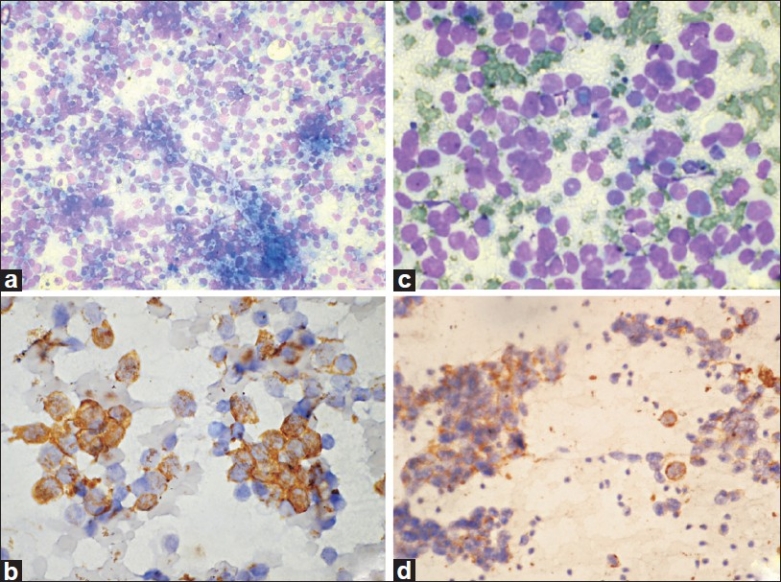
Ewing's sarcoma / PNET. (a) Cellular smears with dispersed monomorphic cells in a vacuolated tigroid background (MGG, ×100); (b) Cells show CD99/MIC2 membrane positivity (IHC, ×400). Rhabdomyosarcoma. (c) Undifferentiated tumor cells with scanty to moderate cytoplasm (MGG, ×400); (d) Smears show desmin positivity (IHC, ×200)