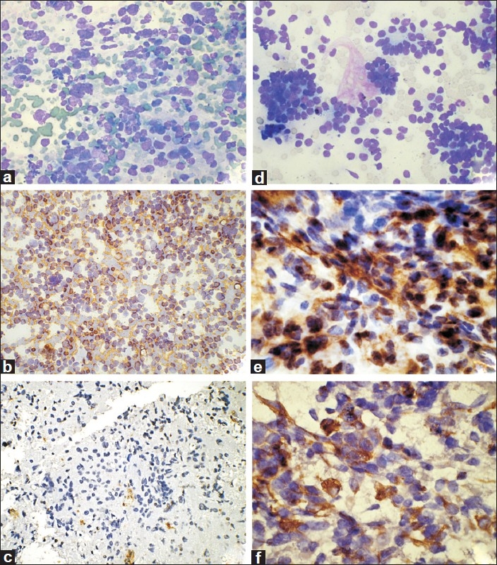
Figure 2.

Neuroblastoma. (a) Cellular smears with dispersed undifferentiated cells (MGG, ×200); (b) Tumor cells are positive for NSE (IHC, 100); (c) Tumor cells are negative for vimentin (IHC, ×100). Wilms’ tumor. (d) Smears showing undifferentiated blastemal cells with focal tubule formation (MGG, ×200); (e) Tumor cells are positive for vimentin (IHC, ×400); (f) Tumor cells are positive for cytokeratin (IHC, ×400)
