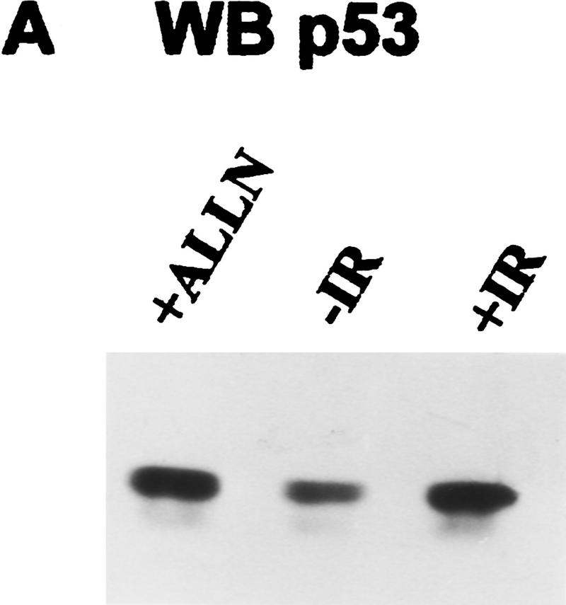
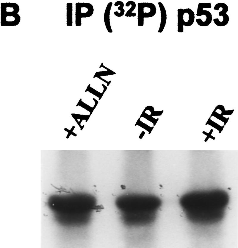
Figure 1.
Immunodetection of p53 protein in ML-1 cells. (A) Western blot analysis of ML-1 lysates. ML-1 cells were untreated (−IR) or treated with either 20 μm of ALLN (+ALLN) or 2 Gy irradiation (+IR). Lysates (50 μg) from each sample were resolved by 10% SDS-PAGE and then electrophoretically transferred to nitrocellulose. p53 was detected by immunoblotting. (B) Immunoprecipitation of p53. 32P-Labeled ML-1 extracts prepared from untreated (−IR), irradiated (+IR), or ALLN-treated (+ALLN) cells were immunoprecipitated with anti-p53 antibodies. Immunoprecipitates were resolved by 10% SDS-PAGE and electrophoretically transferred to a PVDF membrane. Radiolabeled p53 was detected by autoradiography. 32P-Labeled p53 from ALLN- and IR-treated cells had approximately twice as many counts per minute as 32P-labeled p53 from unirradiated cells when the isolated bands were counted in a scintillation counter.


