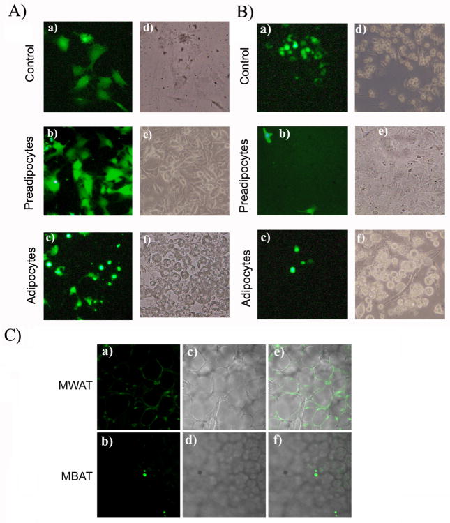
Fig. 2. Change in C2D macrophage cell morphology during co-cultured with adipocytes or pre-adipocytes in vitro and after infiltration into BAT or WAT in vivo.
A) C2D macrophage cells were labeled with CFDA-SE or B) C2D macrophage cells labeled with CFDA-SE and isolated from peritoneal cavity (PEC-C2D) were cultured a) alone or co-cultured with b) 3T3L1 pre-adipocytes or c) adipocytes as described in the Materials and Methods. Panels a, b and c; Cells viewed on the fluorescent microscope (Magnification × 200). Panels d, e and f are phase contrast images of cells in a, b and c. C) WAT-C2D and BAT-C2D were collected from mice two days after adoptive transfer. C2 D macrophages, WAT and BAT were processed as described in Materials and Methods. Panels a and c images from the confocal microscope (× 100). Panels b and d are phase contrast images of the same fields.

