
Figure 6.
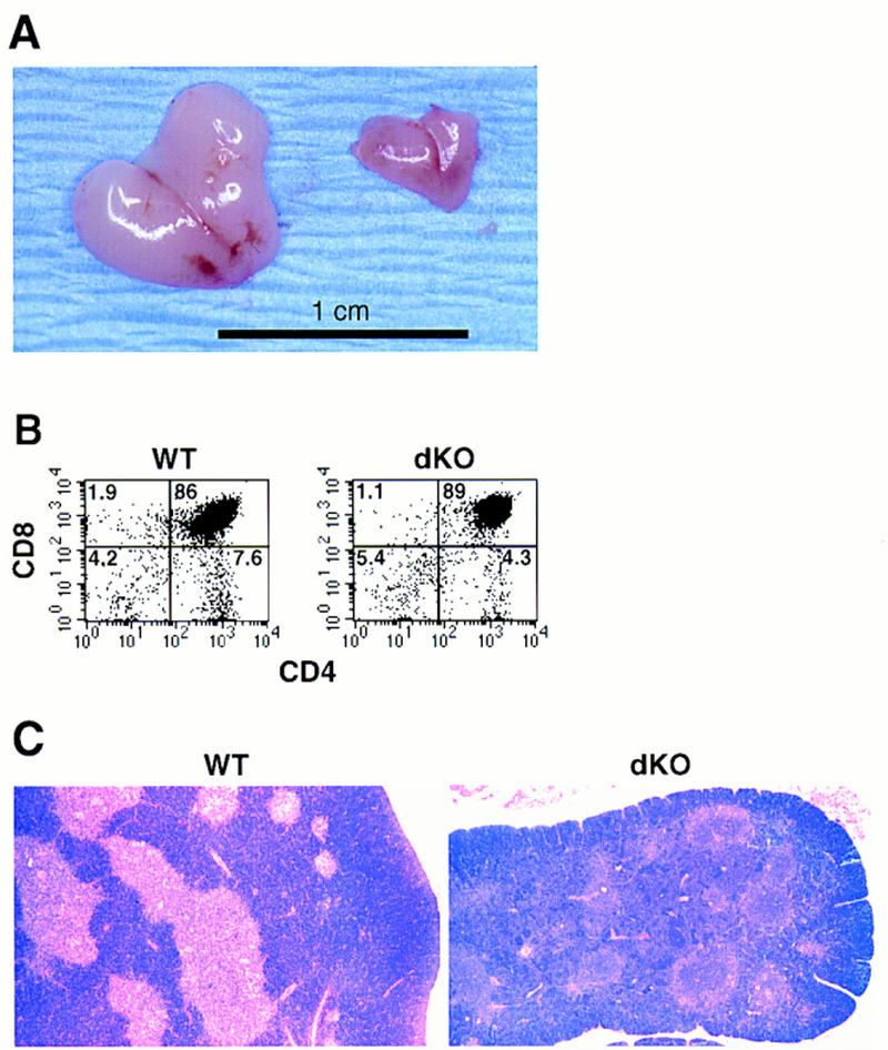
Impaired thymus glands in double knockout mice. (A) Macroscopic appearance of thymic glands obtained from a pair of 17-day-old littermates: (right) double knockout; (left) p50(+/+), p52(+/−). Original magnification was 1.3×. The dKO thymus shown is one of the larger ones seen in mutant mice. (B) FCM analysis of a thymic cell suspension from a 5-day-old double knockout mouse (dKO, right panel) and a littermate control (p50(+/+), p52(+/−); WT, left panel). CD4–biotin vs. CD8a–PE two-color profiles are displayed. Results were confirmed with two additional pairs of mice. Numbers in the quadrants reflect the percentage of total cells in that quadrant. (C) Altered microarchitecture in thymic glands of double knockout mice. Representative, Bouin’s-fixed paraffin-embedded sections were obtained from thymic glands of a 10-day-old double knockout mouse (dKO, right) and a wild-type littermate control (WT, left) and stained with HE, as indicated.