
Fig. 10. Likely spatio-temporal cellular interactions between related CT, TC and TRN neurons during absence-related-SWD in GAERS.
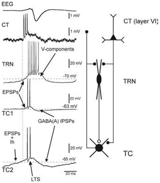
In both non-epileptic control rats and GAERS, at least two types of TC neurons coexist, one of which (TC2) is endowed with a presumed H-current. Note that thalamic, relay and reticular, discharges occur in synchronous, phase-locked manners during the occurrence of an absence-related rhythmic spike-and-wave complex in the cortical surface EEG. Left: SW-related extracellular CT and intracellular TC and TRN activities. From top to bottom: A SW complex (EEG), an extracellular CT discharge, an intracellular TRN discharge, and two typical intracellular TC1 and TC2 discharges. The second TC cell (TC2) exhibits a presumed H-current that coincides with an EPSP barrage. The ramp-shaped depolarization, which includes a presumed Ih, can trigger a low-threshold Ca2+ spike (LTS). In TRN cells, the EPSP barrage can trigger voltage-dependent components (V-components, including a low-threshold Ca2+ potential). Right: Schematic of the anatomical relationships between the three main elements that make up the TC system. Adapted from (Pinault, 2003).