
Fig. 13. Triple extracellular field potential recordings of somatosensory-related thalamic and cortical sites along with spontaneously occurring SWDs in the surface EEG.
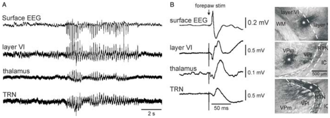
The photomicrographs in B (up, dorsal; right, lateral) show large extracellular applications (epicentre indicated by a white asterisk) of dextran biotin amine where extracellular field potential recordings were carried out simultaneously in the somatosensory system, that is, from top to bottom, in layer VI, in the thalamus, and in the thalamic reticular nucleus (TRN). The bandpass was 0.1–6 kHz. (B) Left: evoked potentials following electrical stimulation of the contralateral forepaw. Abbreviations: IC, internal capsule; VPl, ventral posterolateral thalamic nucleus; VPm, ventral posteromedial thalamic nucleus; WM, white matter. Adapted from (Pinault, 2003).