
Fig. 15. Overview of the corticocortical (black arrows) and corticothalamic (gray arrows) interdependencies during spontaneous absence seizures in WAG/Rij rats as established by nonlinear association analyses.
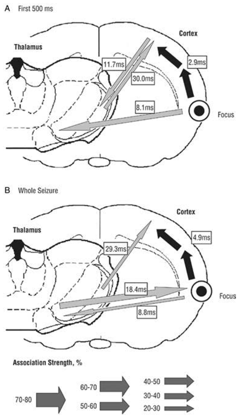
The thickness of the arrows represents the strength of the association, while the direction of the arrowhead points toward the lagging site. (A) The first 500 msec of the seizure. A cortical focus was found in the upper lip and nose area (perioral region, parietal 2) of the somatosensory cortex, as this site consistently led the other cortical recording sites. The hind paw area was found to lag by 2.9 msec with respect to this focal site. Concerning the corticothalamic interrelationships, the cortical focus led the thalamic ventroposterior medial nucleus with a delay of 8.1msec. (B) The whole seizure. The same cortical focus as during the first 500msec was found consistently. Compared with the first 500msec, the time delay from the cortical focus to the nonfocal cortical sites increases and the direction of the corticothalamic couplings changes. Adapted from (Meeren et al., 2005).