
Fig. 6. Normal- and absence-related intracellular SM oscillations in thalamic reticular nucleus neurons are qualitatively similar.
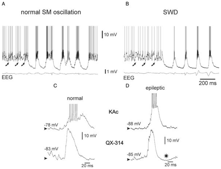
Intracellular recordings of thalamic reticular nucleus neurons during a spontaneously occurring SM oscillation (A) and a spike-and-wave discharge (SWD) (B) in the EEG in a NEC rat and in a GAERS, respectively. The curved arrows in A and B indicate rhythmic barrages of EPSPs. (C,D) Comparison of two typical recurring threshold depolarizing waves, which were recorded during normal SM oscillations in a NEC rat (C) and during SWDs in a GAERS (D), respectively. The intracellular recordings were made with a KAc-(upper traces) or QX-314-filled (lower traces) micropipette. * in D indicates a late hyperpolarization that probably results from a Ca2+-activated K+ current. The action potentials are truncated in C and D for clarity. Adapted from (Pinault, 2003).