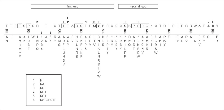
Figure 2.

Mutations in the HBsAg a determinant. The numbers above the line represent amino acid positions in HBsAg. The bold i's between the numbers 120 and 125 represent known insertion points in the surface antigen. Above the numbers is the wild type sequence for HBsAg, with multiple letters in a column denoting alternative amino acids seen in various subtypes. Emboldened positions denote putative residues that participate in the a determinant. The lower case n at position 146 is the site of N-linked glycosylation. Boxed positions show the positions at which mutations are most frequently seen. Below the line the columns of letters represent the mutant residues seen at each position. Asterisks denote stop codons. Numbers below the bold i's denote the oligopeptides inserted at these two positions. These oligopeptides are listed in the box at the bottom. The data for this figure come from several sources.[85,86,89,91,93–95,98,108,110–153]
