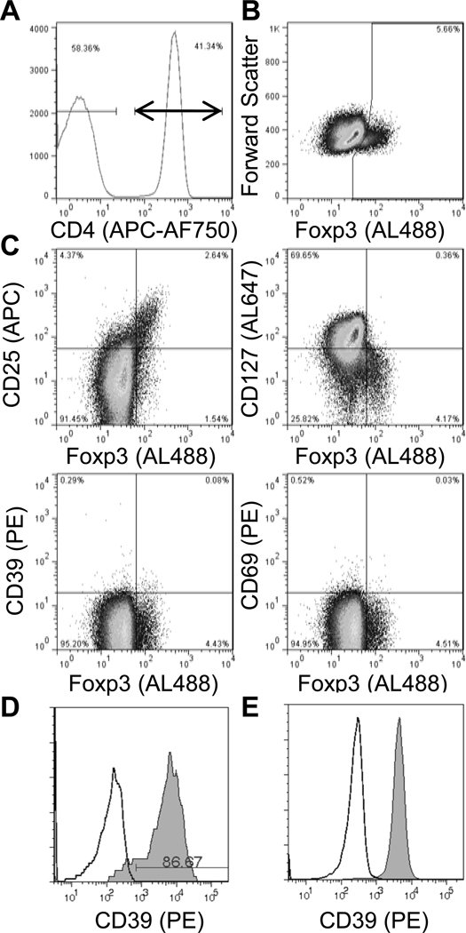
Fig. 2. Lack of CD39 expression in Treg cells from SLE patient.
Freshly isolated PBMC were stained for the indicated surface markers and Foxp3 and then analyzed by flow cytometry. Within gated CD3+ CD4+ lymphocytes (A), Foxp3 expression was analyzed vs. forward scatter (B), or CD25, CD127, CD39, and CD69 (C). Gates were drawn based on isotype control staining (not shown). CD39 expression (shaded; isotype control, open) is shown for gated CD19+ B lymphocytes (D) and monocytes (E).

