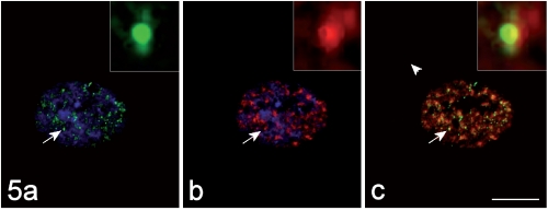
Figure 5.
The immunolabelling for MBNL1 (green fluorescence in a) and that for the snRNP Sm antigen (red fluorescence in b) co-locate (yellow fluorescence in the merged image, c). DNA was counterstained with Hoechst 33258 (blue fluorescence in a, b). Bar: 10 µm. In the insets, the co-labelled focus indicated by the arrow is shown at a higher magnification.

