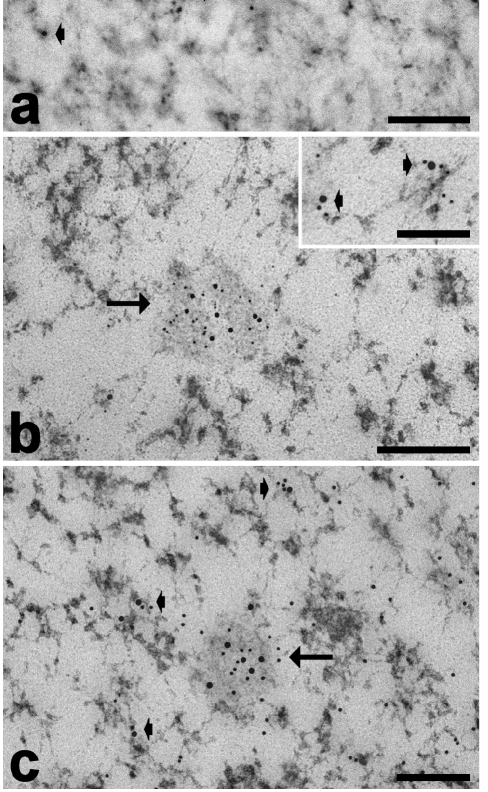
Figure 7.
Myoblast nuclei, paraformaldehyde fixation, Unicryl-embedding, EDTA staining. (a) Immunolabelling with anti-MBNL1 antibody: the signal accumulates in the RNP nuclear domain (arrows) and is also associated with PF (arrowheads). (b) Dual immunolabelling with anti-MBNL1 (12 nm) and anti-snRNP (6 nm) antibodies; (c) dual immunolabelling with anti-MBNL1 (18 nm) and anti-hnRNP (12 nm) antibodies: in both cases the probes co-locate in the RNP nuclear domain (arrow) and on PF (arrowheads). Bars: 250 nm (a–c); 100 nm (insets).

