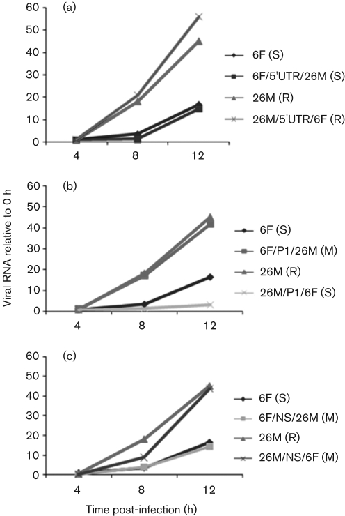
Fig. 7.
Analysis of viral positive-strand RNA synthesis during infection of RD cells. (a) RNA synthesis of parental virus and 5′UTR chimeras; (b) RNA synthesis of parental virus and P1 region chimeras; (c) RNA synthesis of parental virus and P2/P3 region chimeras. Monolayers were infected at an m.o.i. of 1. Culture supernatants were collected at the times indicated. Accumulation of positive-sense viral RNA during infection was measured by quantitative real-time RT-PCR. Yields of viral RNA at each time point were normalized to the yield at the first time point (0 h post-infection). Results represent means of duplicate samples. The growth phenotypes of each chimeric virus are shown in parentheses (S, slow; M, medium; R, rapid).

