
Figure 4.
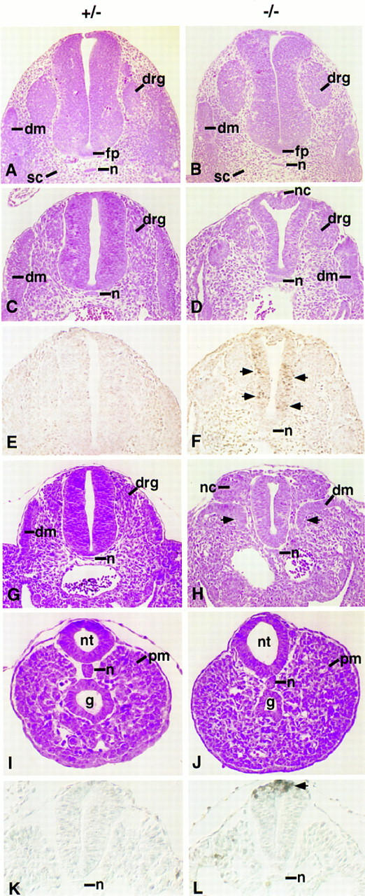
Histological and cell death analysis of spinal cord and somite development in Noggin mutants. Transverse sections through embryos heterozygous (A,C,E,G,I,K) or homozygous (B,D,F,H,J,L) for the targeted Noggin allele at 10.5 dpc (A–J) and 9.0 dpc (K,L). Sections were cut at the level of the forelimb (A,B), between the fore- and hindlimbs (C–F,K,L), at the caudal hindlimb level (G,H), and through the presomitic mesoderm just anterior to the tail bud (I,J). A–D and G–J are hematoxylin and eosin stained; E,F,K, and L underwent the TUNEL reaction to visualize apoptotic cell death. Pairwise comparisons were photographed at the same magnification. (dm) Dermomyotome; (drg) dorsal root ganglia; (fp) floor plate; (g) gut; (n) notochord; (nc) neural crest; (nt) neural tube; (pm) presomitic mesoderm; (sc) sclerotome.