
Figure 3.
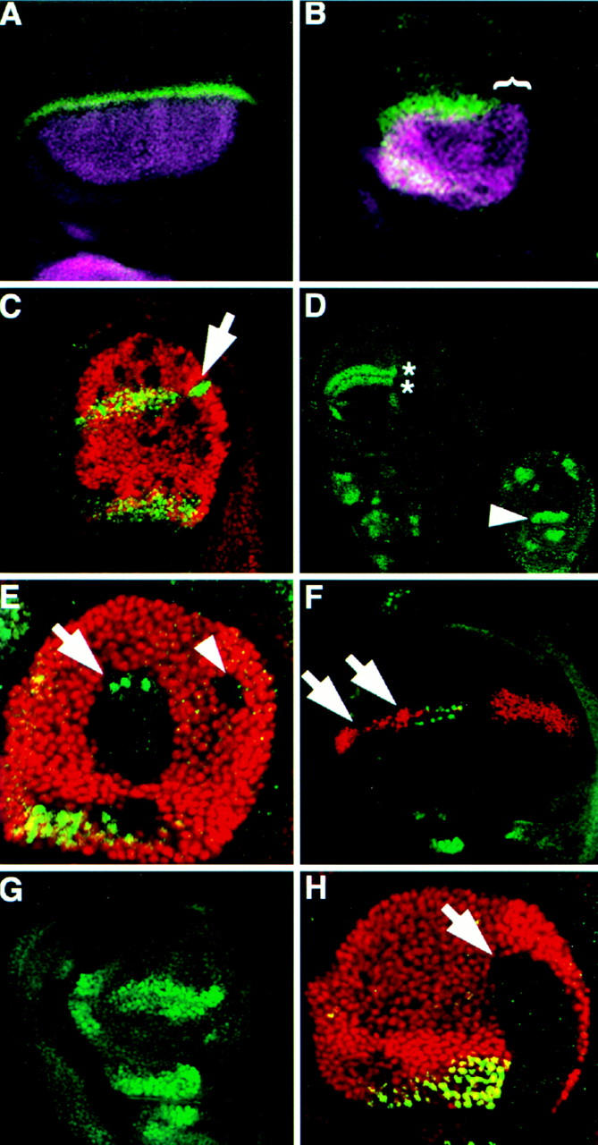
Ubx represses selected genes along the DV boundary of the haltere disc. (A–C) Antibody staining detecting Wg (green); Ap (purple), Ubx (red). (A,B) Ap and Wg are expressed in a similar domain in the haltere (B) as in the wing (A), but Wg expression is absent from the posterior haltere (bracket). (C) Haltere disc with several Ubx− clones (lack of red staining). A posterior clone, located along the DV boundary (arrow) shows derepression of Wg expression. (D–H) Antibody staining detecting Sc (green); Ubx (red). (D) Wild-type expression of Sc in the wing (left) and haltere (right) disc pouches. The double row of expression in sensory organ precursors along the future wing margin (asterisks) is absent from the haltere disc. In the haltere disc, Sc is also expressed in unique patterns including the pedicellular region (arrowhead). (E) Haltere disc with two dorsal Ubx− clones that each touch the DV boundary. The anterior (arrow) clone shows derepression of Sc expression; the posterior (arrowhead) clone shows no Sc expression as in the posterior of the wing. (F) Ubx expression along the DV boundary of a CbxM1/+ wing disc represses Sc expression along the presumptive anterior wing margin (arrows). (G) Ubx6.28/bx34e haltere disc showing ectopic Sc expression along the anterior DV boundary. (H) Ubx− clone (arrow) crossing into the pedicellar region of the haltere disc (see arrowhead in D) fails to activate the normal Sc expression there indicating that Ubx is necessary for activation of Sc in the pedicellar region of the haltere.