
Fig. 4.
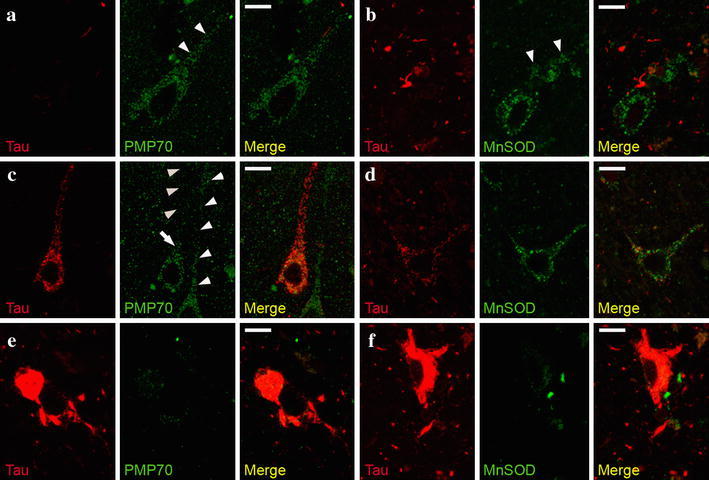
Peroxisomes but not mitochondria are absent from neuronal processes featuring pre-tangles. Representative images of confocal double-immunofluorescence using AT8 antibody to detect phospho-tau (pre-tangle marker) and either PMP70-positive peroxisomes (a, c, e) or SOD2-positive mitochondria (b, d, f) in entorhinal cortex of an AD patient with Braak stage IV. a , b In the absence of phosphorylated tau, numerous peroxisomes (a) and mitochondria (b) are present in the neuronal processes. When phosphorylated tau is present, peroxisomes are only visible in the soma (arrow), but not in the neuronal process (c arrowheads pointing upwards), but normal amounts of mitochondria are present (d). Note the abundance of peroxisomes in a neighboring neuron, in which no phospho-tau is detected (c arrowheads pointing downwards). e, f In neurons filled with NFT, no peroxisomes and only sporadic mitochondria were detected in the soma. Size bar 10 μm