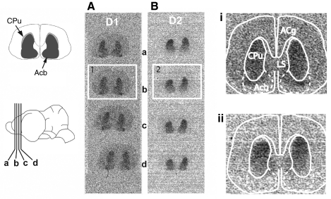
Fig. 3.
Autoradiographic localization of D1- and D2-type dopamine receptors in the Mexican free-tailed bat brain. (A) Representative autoradiograph showing binding in the striatum in four adjacent brain sections (shown inset on the bottom left) incubated with 3H-SCH23390, a D1-type receptor antagonist. (B) Binding in the striatum for four adjacent brain sections incubated with 3H-raclopride, a D2-type receptor antagonist. Panels i and ii are enlarged to the right and overlaid with approximate boundaries of the caudate/putamen (CPu) and nucleus accumbens (Acb) based on Cresyl Violet-stained adjacent sections (shown inset on the top left). LS, lateral septum; Acg, anterior cingulate cortex.

