
Figure 1.
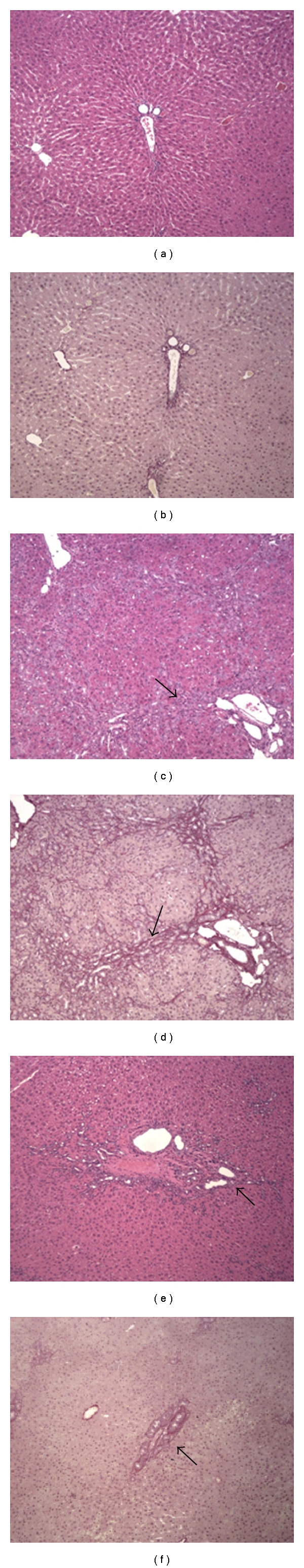
Photomicrographs of liver sections from rats (100x). Liver sections were stained with hematoxylin and eosin (a, c and e) or with picrosírius (b, d and f). (a) and (b): control rats (CO), showing normal architecture of liver parenchyma. (c) and (d): untreated rats with common bile duct obstruction (CBDL), showing regenerative nodules (arrow (c)) and fibrosis (arrow (d)). (e) and (f): Biliary obstructed rats treated with quercetin (CBDL + Q), showing the regeneration of liver parenchyma (arrow (e)) and light fibrosis (arrow (f)).