
Fig. 10.
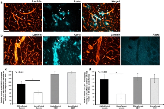
Vascular basement membrane abnormalities in mid and late-stage TG arcAβ mice. Constrictions/stenoses (a, plus signs) and localised thickening of the vessel walls (a, asterisks) were abundantly seen on CAA-laden vessels in brains of late-stage TG arcAβ mice. These basement membrane irregularities due to laminin over-expression (a) were confined to those parts of the cerebral vessels that were affected by CAA; all other parts showed normal laminin expression and smooth vessel wall appearance (a, arrows). Severe CAA in late-stage TG arcAβ mice (b) caused both intense and significant increase in laminin immunopositivity and rupture of the vascular basement membrane laminin (b, arrow; c and d). In addition, dust-like laminin particles could be observed surrounding the affected vessel (b, asterisk). Cerebral vessels of arcAβ NTG littermates lacked CAA and vascular basement membranes showed typical laminin immunopositivity (b, last two image sets). Scale bar a 150 μm; b 30 μm. Hue settings of fluorescence for Aβ were altered for more contrast effect explaining the blue colour