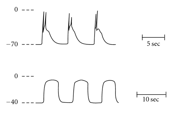
Figure 1.

Voltage profile of electrical activity in cat small intestinal smooth muscle. Shown in the upper panel are spontaneous slow waves and spikes recorded with an intracellular microelectrode. Three slow waves are shown with spikes triggered by the depolarization associated with the upstroke of the slow wave. In the lower panel, prolonged potentials are shown following incubation in calcium-free solution. Membrane potential depolarizes from approximately −70 mV to −40 mV and the voltage excursion of the prolonged potential approaches 0 mV. Note change in time scale between the traces. From reference [9].
