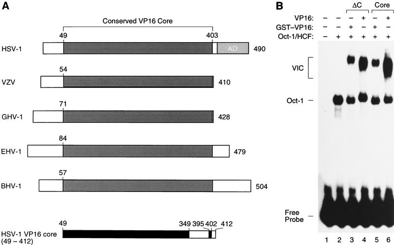
Figure 1.
Conserved VP16-core region. (A) Schematic representation of VP16 homologs from five herpesviruses and delineation of the VP16-core protein used for crystallography. The VP16 homologs are derived from HSV-1 (strain 17; Campbell et al. 1984; Dalrymple et al. 1985), VZV (Davison and Scott 1986), GHV-1 (Yanagida et al. 1993), EHV-1 (Telford et al. 1992), and BHV-1 (Carpenter and Misra 1992). A conserved VP16-core region is shown in dark gray and the carboxy-terminal HSV-1 transcriptional activation domain (AD) is shown in light gray. The HSV-1 VP16 core (49–412) protein is shown at the bottom with the structured regions identified by x-ray diffraction shown in black. (B) Electrophoretic mobility retardation assay of GST–VP16 fusion proteins synthesized in E. coli. A labeled (OCTA+)TAATGARAT site from the HSV-1 ICP0 promoter (Stern et al. 1989) was used for electrophoretic mobility retardation as described (Huang and Herr 1996). (Lane 1) Free probe; (lanes 2–6), partially purified HeLa-cell Oct-1 and HCF (the wheat germ agglutinin fraction; Wilson et al. 1993); (lane 3) GST–VP16ΔC; (lane 4) VP16ΔC; (lane 5) GST–VP1649–412 core; (lane 6) VP1649–412 core. The positions of the free probe, Oct-1–DNA (Oct-1) complex, and VP16-induced complex (VIC) are indicated on the left.

