
Figure 8.
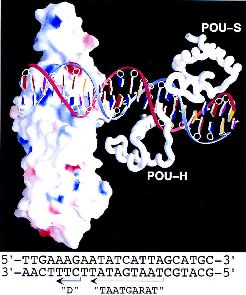
Model of VP16 bound to a TAATGARAT element with the Oct-1 POU domain. The presentation of VP16 is similar to that shown in Fig. 7B. The nucleotide sequence of the DNA in the model is shown at the bottom and originates from the HSV ICP0 gene promoter. In the graphic, the bases are color coded as follows: A (red), T (blue), G (yellow), and C (purple). The blue DNA strand represents the top strand; the red strand represents the bottom strand of the sequence shown below. The Oct-1 POUS (POU-S) and POUH (POU-H) domain structures and the Oct-1 DNA-binding site structure are from Klemm et al. (1994).