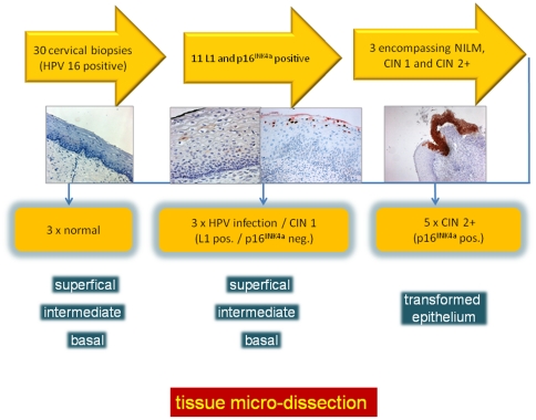
Figure 1. Schematic outline of the recruitment of lesions included into the study.
34 HPV 16 infected cervical lesions were recruited from a gynecopathology archive of cervical punch and cone biopsies. Sections of these lesions were stained with H&E, p16INK4a and L1. 11 of these displayed koilocytes and L1 positive superficial squamous epithelial cells. Of these lesions, three showed a continuum of p16INK4a-positive epithelial areas indicating transforming HPV infections. 2 additional biopsies displaying p16INK4a-positive epithelial areas were recruited from other biopsies not directly showing this continuum of intra-lesional progression for the in depth methylation analysis as shown in Figure 5.

