
Figure 4.
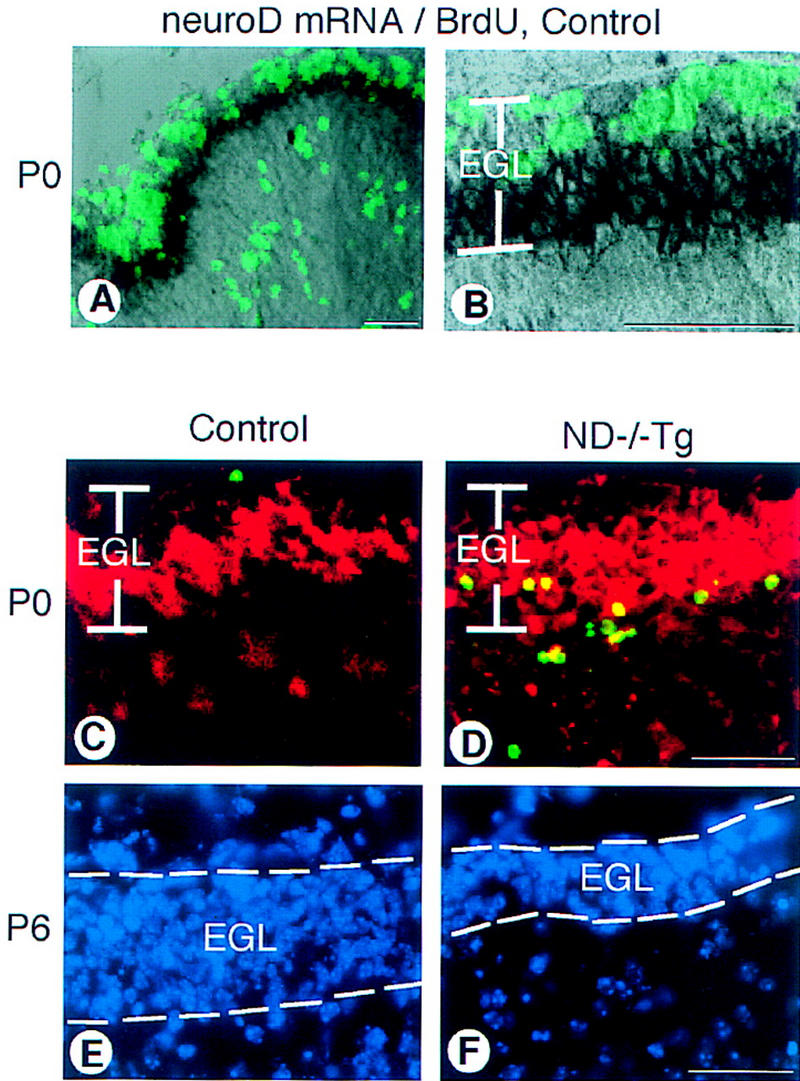
Cerebellar abnormalities in the ND−/−Tg mice. (A) neuroD in situ hybridization with BrdU labeling on control mice at P0. neuroD mRNA (black) is expressed in the inner zone of the EGL; the outer zone is cells incorporating BrdU (green). (B) A higher magnification of the EGL shown in A. (C,D) Apoptotic cells in the P0 cerebellar cortex. In the posterior lobules of ND−/−Tg mice, many TUNEL+ cells (green) are found in the inner EGL where immunostaining with antibody to β-galactosidase (red) is also found. (E,F) DAPI staining shows the reduced EGL thickness in the posterior lobules of ND−/−Tg cerebellum at P6. Bars, 20 μm.