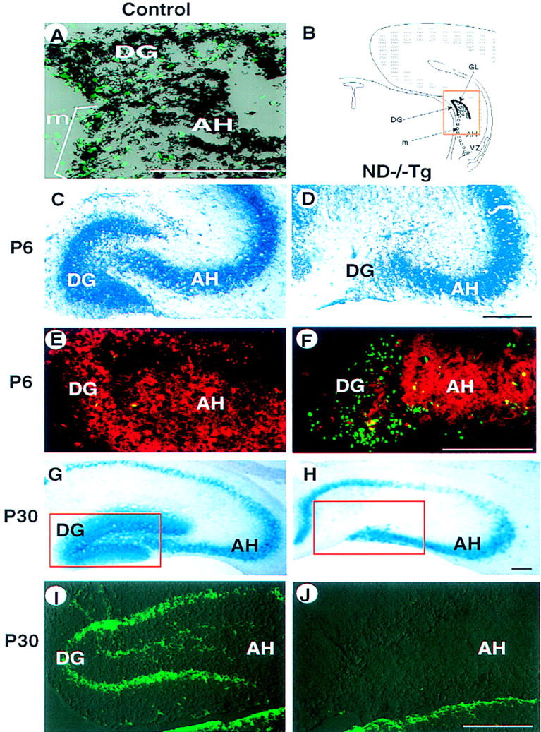
Figure 5.

Hippocampal abnormalities in ND−/−Tg mice. (A) In situ hybridization at P0 showing that neuroD mRNA is expressed (black) in DG-forming granule cells and progenitor cells that are labeled with BrdU (green). (B) Schematic presentation of development of the DG in normal mice. The DG is formed by the granule neurons (●) generated by progenitor cells (○) after migration (m) from the VZ along the AH. (C–J) Degeneration of DG in ND−/−Tg mice as shown by staining with toluidine blue (C,D), anti-β-gal immunostaining (red in E,F), X-gal staining (G,H), or anti-calretinin immunostaining (I and J, corresponding to the squared areas in G and H, respectively; staining in the thalamic area below is also shown) at P6 (C–F) or P30 (G–J). TUNEL staining at P6 (E,F) shows many apoptotic cells (green) in the poorly formed DG in ND−/−Tg mice. (B–F) The squared region in B. Bars, 100 μm.
