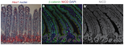
Figure 2. The Notch signaling pathway is activated in the crypts.
(A) In situ hybridisation showing expression of Hes1 mRNA (red silver grains; radioactive probe detection, false color). (B) Immunofluorescence staining for NICD (red), with β-catenin in green and DAPI in blue. (B′) shows the NICD channel only. Note that NICD and Hes1 mRNA are largely confined to the crypts, implying that the crypts are the site of Delta-Notch signaling. The nuclear dots of NICD immunostaining resemble those seen in other studies [55], [56]; a caveat is that this staining may reveal NICD lingering in degradation bodies and not purely the NICD that is active as a transcription factor. Perdurance of NICD immunoreactivity may explain why we saw nuclear NICD staining in a substantial proportion of crypt secretory cells as well as in their non-secretory neighbours (data not shown).

