
Figure 1.
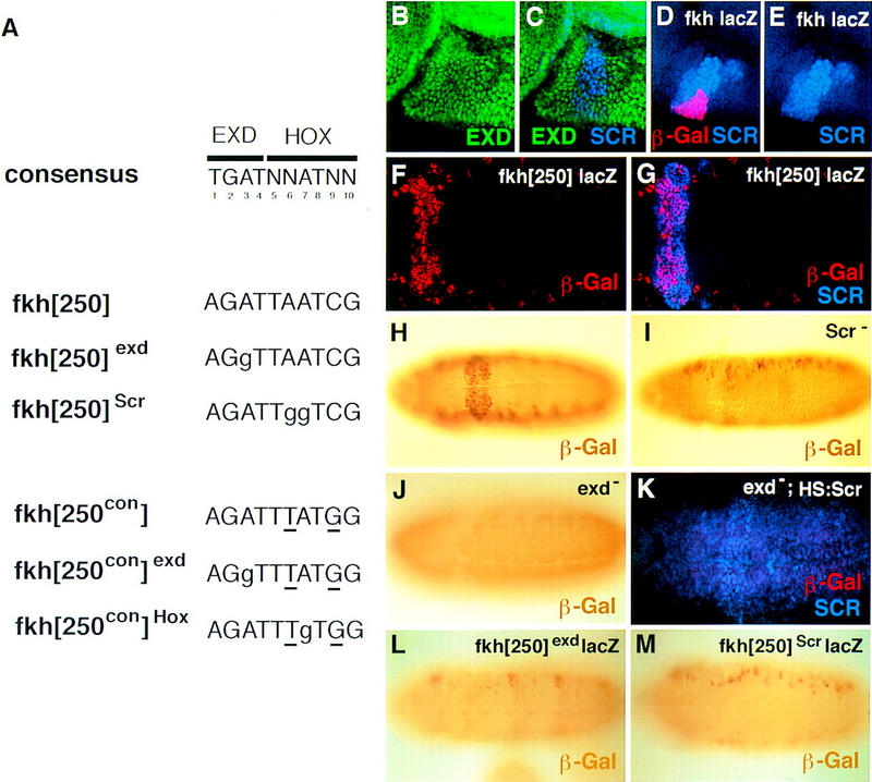
The fkh[250] element includes a potential Hox/Exd-binding site and requires Scr and exd for its activation in embryos. (A) The sequence of the Hox/Exd-binding sites within fkh[250] and fkh[250con] are compared with the consensus-binding site. The two differences between fkh[250] and fkh[250con] (basepairs 6 and 9) are underlined, whereas mutations in the Exd or Hox half-sites are indicated with lowercase letters. (B,C) Fluorescent images of a stage-10 embryo (lateral view of PS 2 region) stained for Exd (green) and Scr (blue). (D,E) Fluorescent images of a stage-10 fkh–lacZ embryo (lateral view of PS 2 region) stained for β-gal (red) and Scr (blue). At this stage, Exd is nuclear in all Scr+ cells, and a subset of Scr+ cells begin to express fkh–lacZ. (F,G) Fluorescent images of a wild-type fkh[250]–lacZ embryo stained for β-gal (red) and Scr (blue). Only a subset of the Scr+ cells express fkh[250]–lacZ. (H,I,J) Wild-type (H), Scr− (I), and exdmat−,zyg− (J) fkh[250]–lacZ embryos histochemically stained for β-gal (brown). PS 2 expression is absent in the Scr− embryo but the lateral staining remains. All detectable reporter gene expression is absent in the exdmat−,zyg− embryo. (K) Fluorescent image of an exdmat−,zyg−; fkh[250]–lacZ em-bryo in which Scr was ubiquitously ex-pressed, and stained for β-gal (red) and Scr (blue). Ectopic Scr was not able to activate fkh[250]–lacZ in these embryos. (L,M) fkh[250]exd–lacZ (L) or fkh[250]Scr–lacZ (M) embryos histochemically stained for β-gal (brown). Mutation of the Exd half-site (L) or Scr half-site (M) abolishes PS 2 expression. (F–M) Ventral views of ∼stage 10 embryos, with anterior to the left.