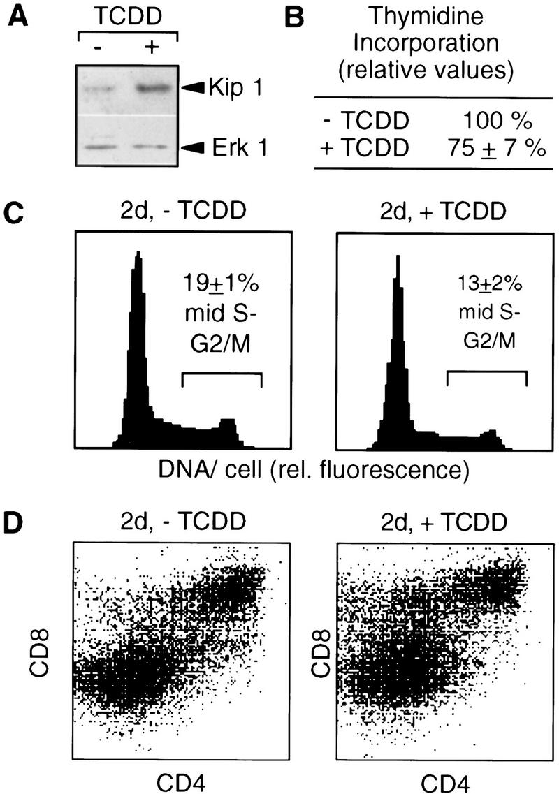
Figure 7.

TCDD-induced p27Kip1 protein and cell cycle delay in FTOCs. Fetal thymus glands were cultured for two days in the presence of TCDD or the DMSO solvent before preparation of thymocytes. (A) p27Kip1 protein in thymocytes was determined by Western blot analysis and even loading was confirmed by reprobing with an Erk1 antibody. (B) Incorporation of [3H]thymidine was quantitated per amount of genomic thymocyte DNA after addition of [3H]thymidine for the last 12 hr of culture. (C) The cell cycle distribution of total thymocytes after 2 days of culture was determined by flow cytometry after DNA staining with H33258. (D) Thymocyte subpopulations after 2 days of culture were analyzed by cell surface staining and flow cytometry. One representative out of three experiments is shown; the numbers in B and C are means ± s.d. from three independent experiments (P < 0.01).
