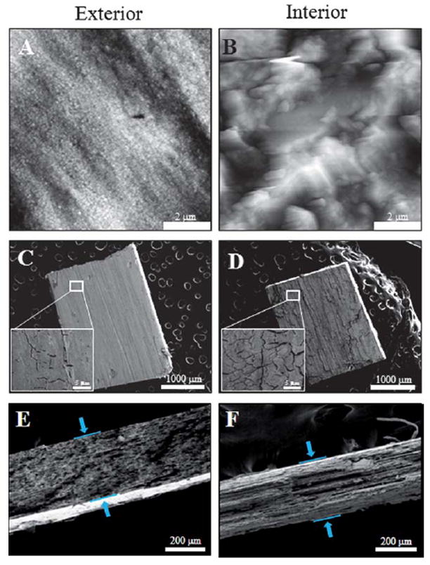
Fig. 6.

Morphology of nanoporous gold. (A) 7 μm × 7 μm, nanoporous gold before peeling, (B) after peeling and captured using AFM. (C) and (D) SEM images of the porous gold before and after peeling. The inset in (C) and (D) are 23 μm × 17 μm zoomed in images of the NPG. (E) and (F) are the side view of the porous gold before and after peeling, respectively, revealing the thickness of the gold plates.
