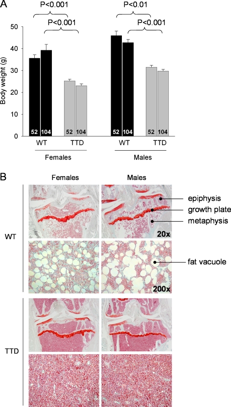
Fig. 2.
a Body weights of female and male WT and TTD, aged 52 and 104 weeks. b Gross histological overview of the proximal tibia of WT and TTD mice, aged 104 weeks. An enlargement (magnification ×200) of the metaphyseal bone marrow is shown below each of the overview pictures (magnification ×20). Please note the absence of fat vacuoles in the bone marrow of TTD mice. This difference was already visible at 52 weeks, albeit less clearly (images not shown)

