
Figure 2.
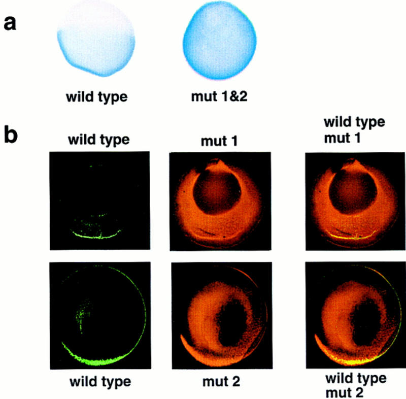
Localization of wild-type and mutant VLEs in oocytes. (a) The distribution of wild-type VLE RNA or double substituted VLE mut 1&2 RNA (1–366/ins1–20&256–275, see Fig. 1a for map) following injection into late stage III oocytes. mut 1&2 RNA appears to be fairly uniformly distributed throughout the oocyte; wild-type VLE shows strong cortical localization to the vegetal hemisphere alone. (b) Confocal micrographs of late stage III oocytes coinjected with wild-type VLE RNA (green channel) and either mut 1 RNA (1–366/ins1–20, see Fig. 1a) or mut 2 RNA (1–366/ins256–275) (red channel). Although the majority of both the mutant RNAs is not localized, some colocalization with the wild-type VLE RNA (yellow, third column) is observable.