
Figure 2.
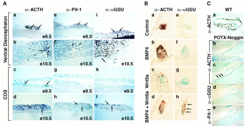
BMP activity is required for anterior pituitary development. (A) Requirement for signaling from the ventral diencephalon in the induction of pituitary lineages. (a,e,i) Coculturing of E9.5 ventral diencephalon with E9.5 Rathke’s pouch explant for 6 days resulted in the appearance of all major lineages as shown by immunohistochemical staining with ACTH (a), Pit-1 (e), and αGSU (i) antisera. (b,f,j) Coculturing experiments done under the same conditions with explants from E10.5, respectively, resulted in the same outcome as in a,e, and i). (c,g,k) Coculturing E9.5 Rathke’s pouch explants in the presence of COS cells for 6 days led to the appearance of only few ACTH-positive cells (arrows, c), no Pit-1 and αGSU-positive cells were detected (g,k). (d,h,l) Coculturing experiment done under the same conditions as under (c,g,and k) with Rathke’s pouch explants from E10.5, resulting in the appearance of ACTH (d)-, Pit-1 (h)-, and αGSU (l)-positive cells. Results are shown from n = 5 independent experiments. (B) Synergistic induction of αGSU expression through BMP and Wnt signaling. (a,e) E9.5 Rathke’s pouch explants cultured in the presence of COS cells showed induction of only ACTH expression (a) as revealed by immunohistochemistry; no αGSU expression (e) is detectable. (b,c,f,g) Similar results were obtained as in a and e when E9.5 Rathke’s pouch explants were cultured with COS cells secreting either BMP4 or Wnt5a alone. (d,h) Coculturing of COS cells producing both BMP4 and Wnt5a with E9.5 Rathke’s pouch explants resulted in induction of αGSU expression (h). Results are from n = 4 independent experiments. (C) Expression of a POTX–Noggin transgene leads to an arrest of pituitary development. (a) Wild-type (WT) littermate at E17.5 stained for ACTH. (b) Phenotypic appearance of the pituitary gland in a POTX-Noggin transgenic embryo at E17.5. The gland has not progressed beyond a single layer epithelium stage reminiscent of Rathke’s pouch at E10.0. A connection towards the oral cavity still remains. (c) Higher magnification of b. Arrows mark the few ACTH-positive cells that can be found in this gland. (d,e) Immunohistochemical staining of adjacent sections with αGSU and Pit-1 antisera, respectively. No positive cells for either of the two markers could be found. Four independent transgene-positive embryos from this stage of gestation were obtained with three showing an abnormal phenotype.